Seattle Children’s Hospital

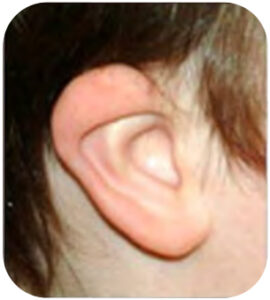
Microtia Reconstruction

Creating ears for kids born without them, providing a more normal and confident sense of themselves. Microtia is the incomplete development and growth of the outer ear. It leads to a small, abnormally-shaped, or absent ear. It occurs with an incidence of 1-10 per 10,000 births and can be divided into four types.

Although associated with many syndromes, microtia typically occurs in isolation and only on one side. The right side is more commonly affected, and males have a 30% higher affected rate than females.

Many children with microtia also have a small jaw on the same side. This is called hemifacial microsomia (HFM) or craniofacial microsomia (CFM). Many children with microtia also have mild abnormalities of their normal ears. About 10% of children with microtia have abnormalities in other systems. These may include facial clefts, heart defects, and eye, kidney, and vertebral abnormalities.

Most of the time we do not know what caused the ear to form abnormally. Exposure to high doses of vitamin A and maternal diabetes during pregnancy are two of the known causes of microtia. There are also some syndromes associated with small ears. These include Treacher-Collins syndrome, Oculoauricolovertebral syndrome, and Goldenhar syndrome. Microtia may run in families, but a specific gene has not been identified yet. Right now there are no tests to show the cause of microtia.

For over the past 20 years, Drs. Kathleen Sie and Craig Murakami have to lead a team that treats microtia patients. Dr. Bhrany is a part of that team along with Drs. Michael Nuar and Randy Bly. The microtia reconstruction program is centered at Seattle Children’s Hospital while adult patients are treated at the University of Washington Medical Center.

Options for the management of microtia include:

  • Observation with no repair
  • Placement of an ear prosthesis
  • Ear reconstruction with either autologous rib cartilage or alloplastic (Medpor) implant
  • After consultation and discussion with the patient and family, a plan that meets both reconstructive and functional goals is chosen. If a microtia reconstruction is chosen, the type of repair is coordinated with hearing management and the possibility of aural atresia repair (creation of an ear canal).

What should we do for our child?

Start by having your child’s hearing tested. We expect that your child will have some hearing loss in the small ear. The hearing test will also tell us about the hearing in the other ear. Children with normal hearing in one ear often develop normal speech and language. However, the presence of hearing loss in one ear can also present some challenges for young children as they learn speech and language. There are two main types of hearing tests: BAER (brainstem auditory evoked responses) and behavioral testing (audiogram). BAER testing is performed if your child is too young to cooperate with behavioral testing. Behavioral testing is done when the child is mature enough to cooperate. Other tests may be recommended, based on your child’s age. Based upon the results of the hearing tests, the audiologist and otolaryngologist will discuss the options for hearing management.

This table provides an outline of our recommendations for your child:

What are the indications for microtia reconstruction?

indications for microtia management should be based on a discussion with the patient and the patient’s family. It is important to understand the patient’s goals and expectations for ear reconstruction. We strive to help patients understand the different options and possible outcomes. A reconstructed ear will make it easier to wear glasses and may permit the patient to use a hearing aid.

The majority of patients with microtia also have aural atresia (absence of an ear canal) associated with hearing loss. Management of conductive hearing loss has implications on microtia reconstruction. All options both for hearing rehabilitation and ear reconstruction are thoroughly discussed with the patient and family. Drs. Sie and Bhrany work with the patient and family to generate a cohesive plan that includes the management of the ear and hearing.

What are the options for hearing for patients with microtia?

HEARING REHABILITATION OPTIONS IN UNILATERAL AURAL ATRESIA

From a surgical planning perspective, one of the main decision points in determining if a patient is an appropriate candidate for the creation of an ear canal (Atresiaplasty). The decision is based on high-resolution computed tomography (CT) of the temporal bones typically done around age 4 years. Obtaining the CT scan at about 4 years of age obviates the need for sedation, allows for mastoid bone growth, and may mitigate the potential effects of radiation on the developing brain. This timing also allows for the CT scan to screen for occult congenital cholesteatoma. Traditionally, autologous cartilage microtia reconstruction was performed prior to atresiaplasty. More recently, multiple surgeons have reported atresiaplasty before or simultaneous with microtia reconstruction with good outcomes.

What are the options for microtia reconstruction?

The only surgical microtia reconstruction option for many years utilized autologous (the patient’s own) rib cartilage, and surgeons preferred to wait until the patient was at least 6 years of age for multiple reasons: (1) the opposite ear is near full size, (2) the rib cartilage is of adequate size, and (3) the patient is able to understand the reconstructive options. The last point could be considered a disadvantage in that families may prefer to undergo the reconstruction as soon as possible and before school begins.

The introduction of alloplastic (Medpor implant)

The advantages and disadvantages of each type of microtia reconstruction option are described below. Drs. Sie and Bhrany have extensive discussions with patients and their families as well as reviewing photographs of patients who have undergone various reconstruction techniques to aid in their decision.

MICROTIA MANAGEMENT OPTIONS

What is the process to undergo microtia reconstruction?

The Consultation

The initial consultation centers around your and your child’s goals of ear reconstruction and an examination of the ears in the context of an overall head and neck examination and hearing management.

During your consultation, options for ear reconstruction including observation with no repair, placement of an ear prosthesis, and ear reconstruction with either autologous rib cartilage or alloplastic (Medpor) implant are discussed. The type of repair is coordinated with hearing management and the possibility of aural atresia repair (creation of an ear canal). A method is chosen that meets both reconstructive and functional goals.

Pre and post-operative examples of other similar microtia reconstructions are reviewed. The details of the procedure and post-operative care are explained, and any questions you may have are answered. If a plan is confirmed to move forward with surgery, a surgery date is scheduled after a discussion with our patient care coordinator.

Pre-operative Visit

Prior to surgery, one more pre-operative visit may be required. This visit typically involves a pre-operative history and physical at Seattle Children’s Hospital for our pediatric patients or the University of Washington Medical Center for adult patients. This visit is performed to assess any medical issues present and to ensure that surgery proceeds in the safest manner. Pre and post-operative instructions are reviewed, and this visit is also an opportunity to ask additional questions that may have arisen after your initial surgery scheduling.

Day of Surgery and Hospital Admission

Once in the pre-operative area, the patient and family will meet the nursing and anesthesiology staff who will take care of you during the procedure. Drs. Sie and Bhrany will meet with you and your family prior to the procedure to review the procedure and discuss any last-minute questions present. After the procedure is performed, the patient will be taken to the recovery room.

A head dressing will be in place as well as a drain tube placed behind the ear. For patients who have undergone first-stage microtia repair with rib cartilage, the drain will remain in place for two days, and the patient will be admitted to the hospital for two days. For patients who have undergone reconstruction with a Medpor implant, the patient will be admitted overnight and the drain removed the next morning.

Post-operative Visits and Follow-up Care

The first post-operative visit will be approximately one to two weeks after surgery to remove dressings and evaluate healing. You will then follow-up likely follow-up at one month after surgery and usually every three months to six months for the first year after your surgery to ensure adequate healing.

What are the risks or complications of microtia reconstruction?

There are risks and complications with microtia reconstruction. Fortunately, the majority of them are minor and easily managed. As with any surgical procedure, there are risks such as bleeding, infection, and risks related to undergoing anesthesia. Issues with wound healing are not uncommon. If wound healing is not ideal, there can be loss of shape of the reconstructed ear with exposed cartilage or Medpor implant. These exposures can result in a less defined ear appearance to even the need for removal of the reconstructed ear and revision reconstruction. Specific to autologous (patient’s own) rib cartilage microtia reconstruction, there is a very low risk of damage to the lining of the lung called the pleura that can result in a pneumothorax (collapsed lung). If the pleura is injured, it is identified and repaired at the time of surgery. There is an approximate 3% risk this injury can result in a pneumothorax (collapsed lung). Rarely, that may require a drain tube to be placed in the chest to help the lungs re-inflate.

With You From Step One

Contact us below or call at 206 599 Face (3223). We’re here to help answer your questions or to get you started with a consultation.